What is ACDF?
In ACDF, a small (approximately 1-inch) incision is made at the front of the neck. The cervical spine is reached by gently dissecting between the tissues without dividing them. This approach allows direct visualization of the disc that is causing nerve compression.
Once the correct disc level is confirmed, the damaged disc is removed under high-powered microscopic magnification, relieving pressure on the nerve and/or spinal cord.
Surgical Approach
A small incision is made at the front of your neck. The surgeon gently retracts the tissues to access the cervical spine, allowing for precise removal of the damaged disc.
Removing the Damaged Disc
Under high-powered microscopic magnification, the damaged disc is carefully removed. This step is crucial as the disc is often the source of nerve compression and pain.
Relieving Nerve Compression
The spinal cord and exiting nerves are visualized and freed from any compression by disc fragments or bone spurs, providing immediate relief.
Inserting a Cage
After disc removal, a spacer (or "cage") is inserted into the disc space. This cage is made of durable metal or plastic and filled with bone graft or a substitute. It maintains disc height and allows the vertebrae to fuse together, forming a stable column.
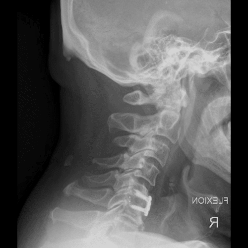
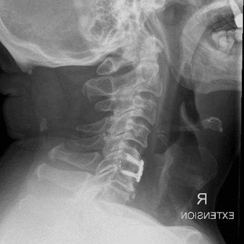
Placement of Plate and/or Screws
To ensure optimal fusion and stability, a low-profile plate and/or screws are used to secure the adjacent vertebrae. These are placed only at the fused level, preserving flexibility at the disc replacement level.
Recovery
Most patients spend one night in the hospital. Recovery times vary, but many experience immediate relief of nerve pain. Minor issues—such as swallowing difficulties and neck tightness—may last 3-4 weeks. A collar may be provided for comfort, and specific postoperative instructions are given by Prof Aaron Buckland’s team.